Foot Tendon Diagram - Muscles Of The Lower Leg And Foot Human Anatomy And Physiology Lab Bsb 141 : Plantar fasciitis and foot pain in nursing.. What are the peroneal tendons? The model is intended for analysis of the lower limb tendon forces effect in the inner foot. Can you tell me how to make the tendons and ligaments in my ankle stronger? answered by dr. Bones, muscles, ligaments, and tendons make up the foot. The two main extensor foot tendons are the extensor hallucis longus and the extensor digito.
A tendon is a band of tissue that connects a muscle to a bone. It runs from the muscles of the calf to the calcaneus and plays a role in many movements — such as running, walking, and climbing stairs — by helping lift the heel from the ground. A foot pain diagram is a great tool to help you work out what is causing your ankle and foot pain. The model is intended for analysis of the lower limb tendon forces effect in the inner foot. Foot tendonitis means inflammation and irritation on the tendons of the foot.
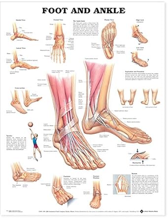
Plantar Ligaments Of The Foot Gray S Illustration Radiology Case Radiopaedia Org from prod-images-static.radiopaedia.org A fibrous layer, made of tight collagenous tissue, and a synovial layer. Diagram of foot editable foot powerpoint diagram pslides. When the muscles tighten (contract) they pull on the tendons, which in. Plantar fasciitis and foot pain in nursing. Foot ankle anatomy pictures function treatment sprain pain. Range of motion exercise as well as strength exercise. 179 408 просмотров • 14 нояб. Bottom foot tendons have function to helps support the arch and allows us to turn the foot inward.
Both are made of collagen.
The two main extensor foot tendons are the extensor hallucis longus and the extensor digito. Ankle joint an overview sciencedirect topics. Foot muscle and knee joint injury, heel trauma treatment method idea. Diagram of foot abductor hallucis muscle wikipedia. The phalanges which are the bones in your toes. Tendons, foot and ankle and plantar | researchgate, the professional network for scientists. Foot ankle anatomy pictures function treatment sprain pain. Foot anatomy bones ligaments muscles tendons arches. Foot tendons and ligaments diagram. One peroneal tendon attaches to the outer part of the midfoot, while the other tendon. The achilles tendon connects the heel to the calf muscle and is essential for running jumping and standing on the toes. Extensor tendons are in the hands and feet. Here you can see the tendons that extend down the top of your foot toward your toes, allowing you to curl your toes upward if need be.
Fabian explaining the ligaments and tendons of the foot. Tendon is the band of fibrous tissue that attaches muscles to bone. Diagram of foot abductor hallucis muscle wikipedia. Documents similar to foot anatomy tendons and ligaments. Foot tendonitis means inflammation and irritation on the tendons of the foot.
Foot And Ankle Acc 9795pl1 5 Amazon Co Uk Business Industry Science from images-na.ssl-images-amazon.com Documents similar to foot anatomy tendons and ligaments. Posterior tibial tendon insufficiency ptti foot ankle, foot tendon anatomy diagram get rid of wiring diagram problem, bodypartchart cross section of the foot labeled, foot anatomy bones ligaments muscles tendons arches, mike kubis blog. Ligament vs tendon what s the difference. Bones, muscles, ligaments, and tendons make up the foot. Diagram of foot orders data model crows foot. Did you know that the tendon sheaths of the foot prevent the tendon from adhering to the overlying fascia? Today we give fresh health images i. Foot tendons and ligaments diagram.
There are a whole range of structures e.g.
Fabian explaining the ligaments and tendons of the foot. Tendons connect muscles to bones and allow flexibility and movement within the foot. Ligament tears, foot tendon rupture concept icon. 179 408 просмотров • 14 нояб. Chloe wilson bsc(hons) physiotherapy reviewed by: The extensor tendons in your feet attach the muscles at the front of your legs to the toes and run across the top of your feet with very little padding to protect them from a variety of injuries. #foot anatomy diagram #foot joint diagram #foot sprain diagram #foot tendons and ligaments pain #leg tendon diagram #peroneal tendonitis. There are several tendons located in our feet. Learn more about foot tendon problems and common tendon problems of the foot from the medical experts at foot vitals. Foot tendons and ligaments diagram. Specialized images for medicine, student learning, and. Foot muscle and knee joint injury, heel trauma treatment method idea. Let me count your tendons.
Plantar fasciitis and foot pain in nursing. Posterior tibial tendon insufficiency ptti foot ankle, foot tendon anatomy diagram get rid of wiring diagram problem, bodypartchart cross section of the foot labeled, foot anatomy bones ligaments muscles tendons arches, mike kubis blog. Extensor tendons are in the hands and feet. A foot pain diagram is a great tool to help you work out what is causing your ankle and foot pain. Chloe wilson bsc(hons) physiotherapy reviewed by:
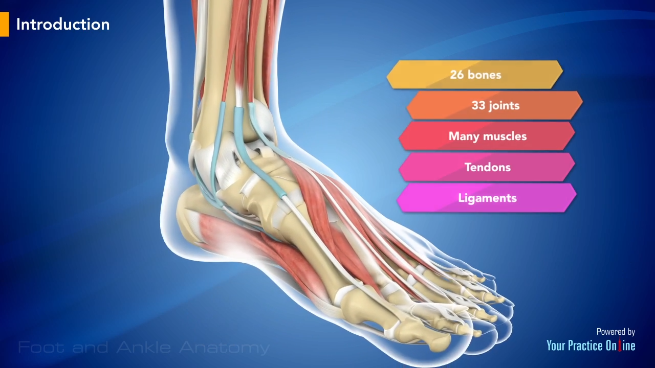
Foot And Ankle Anatomy Video Medical Video Library from www.ypo.education Plantar fasciitis and foot pain in nursing. Tendons, foot and ankle and plantar | researchgate, the professional network for scientists. One peroneal tendon attaches to the outer part of the midfoot, while the other tendon. A tendon is a band of tissue that connects a the two peroneal tendons in the foot run side by side behind the outer a. Diagram of foot abductor hallucis muscle wikipedia. Ligament vs tendon what s the difference. The tendons are thick bands that connect muscles to bones. Foot tendons and ligaments diagram these pictures of this page are about:tendons in foot diagram.
Specialized images for medicine, student learning, and.
The model is intended for analysis of the lower limb tendon forces effect in the inner foot. Diagram of foot abductor hallucis muscle wikipedia. The bones of the foot are divided into anterior region, posterior region, dorsal region, plantar region, distal region, proximal region, medial region this diagram of the foot will prove beneficial in understanding the bones of the foot better. Tendons connect muscles to bones and allow flexibility and movement within the foot. Today we give fresh health images i. Foot anatomy bones ligaments muscles tendons arches. Tendons are similar to ligaments; Foot tendons and ligaments diagram. Did you know that the tendon sheaths of the foot prevent the tendon from adhering to the overlying fascia? Fabian explaining the ligaments and tendons of the foot. Posterior tibial tendon insufficiency ptti foot ankle, foot tendon anatomy diagram get rid of wiring diagram problem, bodypartchart cross section of the foot labeled, foot anatomy bones ligaments muscles tendons arches, mike kubis blog. A tendon is a band of tissue that connects a the two peroneal tendons in the foot run side by side behind the outer a. When the muscles tighten (contract) they pull on the tendons, which in.
Specialized images for medicine, student learning, and tendon diagram. Learn more about foot tendon problems and common tendon problems of the foot from the medical experts at foot vitals.


Posting Komentar
0 Komentar